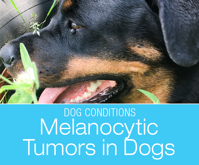
Melanocytoma in a Dog: Cookie’s Eyelid Bump that Was Not a Cyst
A melanocytoma is a benign tumor of cells that produce pigment—melanocytes. Only a histopathologist can determine the difference between a melanocytoma and a malignant melanoma.
As a general rule, tumors of the pigment-producting cells are usually benign where the skin is covered by fur and malignant in bare-skin areas such as lips or nail beds. The one exception are eyelids where these tumors are usually benign as well.
Further information: Melanocytic Tumors
Location, location, location.
In dogs, melanoma isn’t strongly linked to sunlight. Tumor behavior depends less on the pigment cell itself and more on the neighborhood it lives in.
Fur-covered skin
Fur protects against trauma, UV, and microbes. Tumors here tend to grow slowly and stay local.
- thicker, more protective layers
- less irritation and trauma
Hairless or moist inner linings
(lips, mouth, nail beds)
Tumors here are more likely to invade and spread.
- thinner, more permeable tissue
- more inflammatory/immune activity due to bacteria or mechanical trauma
- richer blood supply
Eyelids behave more like haired skin and are partly protected by lashes and surrounding fur. That’s why most eyelid tumors, ~75-90%, turn out harmless. Chronic irritation, inflammation, or invasion of delicate inner tissues can push a tumor toward trouble.
Signs an eyelid bump deserves a closer attention
- growing or changing shape
- rubbing on the eye
- redness, discharge, or squinting
- pawing at the eye
- bleeding or ulceration
- interfering with blinking
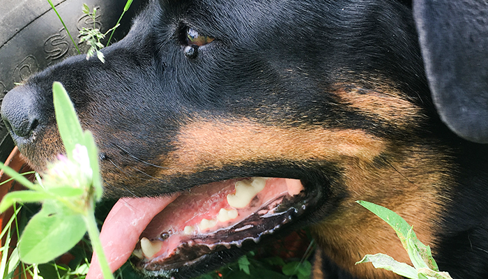
Cookie’s story
A little over a year ago, I noticed a tiny bump on Cookie’s lower eyelid. It was so small that you could only see it at a certain angle and in a certain light. I wasn’t even sure whether I was just imagining things.
I brought it up during her nearest wellness exam to make sure it’s not something to worry about. Cookie’s veterinarian assured me it was nothing dangerous—most likely a cyst. It didn’t make sense to take any drastic measures in trying to get a better identification.
Little bump grows
Very slowly, the tiny bump kept growing. I discussed it with the veterinarian again, this time because I was concerned about it hurting the eye. It was, however, clearly growing in the direction away from the eye. Should Cookie need to go under for another procedure, we would remove it then.

Meanwhile, Cookie saw an orthopedic specialist to figure out why her shifting front leg lameness wasn’t improving. The diagnosis was mild elbow dysplasia, and we chose platelet-rich plasma (PRP) therapy.
Normally this is done under heavy sedation. But:
- the specialist recommended repeat injections to boost the effect
- heavy sedation was a bad option for Cookie
Further information: Front Leg Lameness in a Rottweiler: Cookie’s Sore Front Legs
General anesthesia instead of sedation
To give Cookie her booster PRP, we had a dilemma. The standard sedation protocol caused severe side effects for her. The adjusted version carries a higher risk of a dangerously slow heart rate, especially in Rottweilers.
A carefully planned general anesthesia was the safer choice. Since she’d already be under, we decided to take care of a few things at the same time—remove the eyelid lump and get dental x-rays. So that’s what we did.
Related articles: Rottweiler Anesthesia Sensitivity: Navigating Sedation and Medication Challenges with Your Large-Breed Dog
Removing Cookie’s bump
Cookie’s veterinarian took great care to make everything as safe and smooth as possible. That day, Cookie went under anesthesia, received her PRP injections, had dental x-rays, and the vet removed the bump. The tissue was sent to the lab for analysis.
Up to that point, we still thought it was just a cyst. But better safe than sorry—I never have a lump removed without sending it for testing.
Melanocytoma
While Cookie’s incision was healing, we received the report from the histopathologist—it wasn’t a cyst after all. Fortunately, it was benign and completely removed. Still, the specialist couldn’t resist adding the usual small-print reminder that things can occasionally go sideways.
Canine dermal melanocytic neoplasms with these histologic features are typically benign and complete surgical excision can be curative. In a small percentage of cases, tumors that are histologically benign can display locally infiltrative growth or distant metastasis.
Fabiano Oliveira, DVM, MS
Because the excision was complete, we’re confident Cookie was in the clear. Of course, if anything changed, we’d act quickly—but for the time being, her veterinarian considered this chapter closed.
It’s another reminder: you can’t judge a bump by its cover. If this hadn’t been such a delicate location, I would have aspirated it immediately. Every other lump gets tested—no exceptions.
Eyelid Melanocytoma: Breed Tendencies
Melanocytic eyelid tumors can occur in any dog, but they’re reported more often in:
- Schnauzers (Miniature and Standard)
- Poodles
- Cocker Spaniels
- Labrador Retrievers
- Golden Retrievers
- Doberman Pinschers
Breed may increase the chance of developing a tumor, not the chance of it being malignant. Age matters more than breed—these growths show up most often in middle-aged and senior dogs.
Melanocytoma in a Dog FAQ
No. Most eyelid pigment tumors in dogs are benign melanocytomas. Malignant melanoma is much less common.
No. Many bumps look alike—cysts, warts, gland tumors, melanocytomas, or melanoma.
Only lab analysis (histopathology) gives a definitive answer.
Not always.
Small, stable bumps that aren’t irritating the eye can sometimes be monitored.
Growing, changing, or irritating masses are usually best removed while still small.
Smaller masses are easier to remove cleanly, and less likely to irritate the eye. Removal avoids potential large incision later and you get a diagnosis sooner instead of guessing.
A small percentage can behave aggressively, especially if they invade the inner eyelid or surrounding tissue. That’s why biopsy matters.
Related articles:
What Is That Bump on My Dog: Canine Lumps, Bumps, and Growths
Further reading:
Melanocytic Tumors
They eye is always such a scary area to do anything. I’m glad it wasn’t anything bad for Cookie. My Husky had a lot of little bumps as she aged, but the Vet said none were concerning – she called them “old lady bumps”.
Yes, eyes are a scary area. When Jasmine had a renegade lash–she needed that removed, I was concerned too. Fortunately all eye problems worked out well.
Excellent info, Jana. I’m glad Cookie’s was benign! I’m the same way – I always had any growths removed from my dogs sent off for pathology (which thankfully, were all benign). My Mom’s one Poodle had one of these lumps when she was about 13. Thankfully, it too, was benign. Sharing this info with my FiveSibes readers!
As it turns out, location matters and the eyelid ones typically are benign. Personally, if the growth is large enough, I prefer aspirating before removal because safety margins are important if it turns out malignant.
Super info about what a melancytoma is and what to look for with these growths. It is scary to see a lump and know that it’s growing and can impede your dog’s well-being. Such a great example that having a vet you trust is vital for your buddy’s care.
Yes, a vet one can trust is gold. And in my case, a ton of research too 😉
Good to hear it was benign! Putting a dog under anesthesia is so worrying, I know it was an issue for one of my dogs but I had complete faith in my vet. It’s so important to have new issues whether behavioural or physical checked out, as it often leads to a more positive outcome.
Knowing is always better than guessing.
Unfortunately, both my heart girls had major issues with narcotics whether with anesthesia or otherwise.
Hi, I just had the exact same thing with my rottweiler. She also had 2 bumps that were removed and sent to analysis. The vet just called me and told me that it was melanocytoma and that it even though it was benign it was good to have it removed. May I ask if you have any follow up infos that you can share? Was the problem solved for good? Thank you 🙂 best regards
So far it has not returned; so it seems it is gone for good. I suspect the trigger might have been some of the deer fly bites–they really love going after her eyes.
I am glad to hear that it turned out to be something benign. I know that fear that comes with finding an unexplained lump, we’ve been there with our pets before. Your mind races and all the most negative possibilities start to haunt you. Cookie is lucky to have such a great owner doing everything to keep her happy and healthy!