A Primer on Cytology: When You Might Need One For Your Dog?
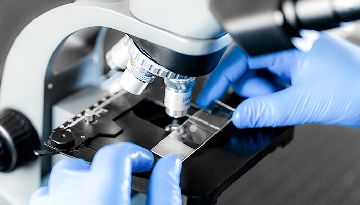
Cytology is the examination of cells under a microscope.
Cytology can help to characterize lumps and masses on the skin and in other organs of your dog. It is also helpful for evaluating:
- cells from body fluids (e.g., urine, joint fluid, fluid in the chest or abdomen)
- body surfaces (such as the ear, skin, eye, mouth, vagina, etc.).

The purpose behind it
Cytology often provides diagnostic information about your dog that can be useful in deciding on a treatment plan.
Lumps and bumps
Your veterinarian can obtain cells for analysis via fine needle aspirate (AFN) or biopsy of the mass. Cytology helps determine whether the mass is malignant or benign. The results inform an appropriate treatment plan.
Further, the obtained cells can be tested for staging and other useful information.
Benign growths can either be left alone or removed. However, such an approach can be risky with cancerous masses. That is because the removal of cancerous masses needs to be complete and with clean margins. Therefore it is essential to identify any mass before removal.
Further analysis of the removed tissue determines what other follow-up treatments might be necessary.

Swabs and skin scrapings
Cytology from a swab or a skin scraping of an infected ear can reveal the presence of:
- mites
- bacteria,
- or yeast infections
This allows for a targeted treatment plan.

Vaginal cytology
Vaginal cytology can help characterize a female dog’s estrous stage, or “heat cycle.” This is useful in determining the best time for breeding.
Urinalysis
Examination of the urinary sediment can reveal the following:
- red and white blood cells
- bacteria,
- or crystals
This is useful in the diagnosis of urinary tract infections or other conditions.
Fine needle aspiration (FNA)
The veterinarian collects cells from a mass or lump with a fine needle.
They insert a sterile needle into the mass and directly withdraw cells from the solid tissue. This procedure can also be used to collect a fluid sample from an organ or body cavity.
Swabs
The veterinarian often swabs ears with a Q-tip. They might also collect skin cells by scraping the skin with a small, sharp blade.
Sometimes, it is possible to press a microscope slide directly on a lump or body part to create a smear for examination.
Fine needle aspiration, swabs, and skin scrapings are generally quick, relatively painless, and noninvasive procedures.
In many cases, cytology can provide a great deal of clinical information and sometimes yield a definitive diagnosis. However, in other cases, cytology can indicate the need for additional, more aggressive testing (e.g., taking a tissue sample for biopsy).
Related articles:
Canine Mast Cell Tumors: JD’s Mast Cell Tumor Diagnostics, Strategy, and Treatment
Further reading:
Cytologic diagnoses that every practicing veterinarian should be able to make